Is the lesion benign and does not require histopathological examination? A quicker and less invasive option is laser removal of moles and skin lesions — no scalpel and no stitches.
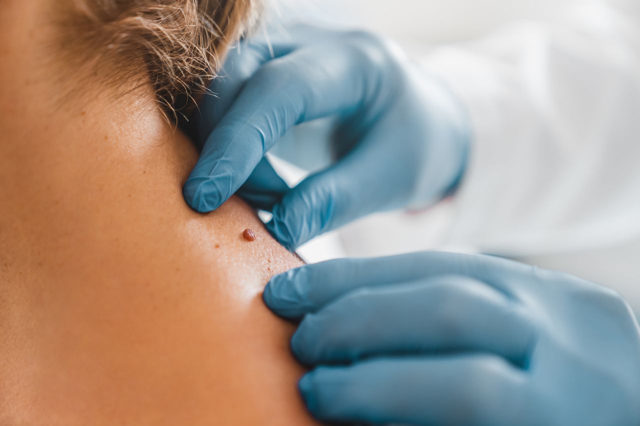
Which moles should worry us? When should you see a specialist?
Types of moles that can be removed surgically at MM Clinic:
Pigmented moles
We choose the surgical method when a mole raises diagnostic doubts and requires histopathological examination, is large, deeply seated or has previously changed in appearance. Excision with a margin of healthy tissue allows the material to be sent for examination — which the laser method does not allow.
Lipomas
A lipoma is a collection of fat cells surrounded by a thin fibrous capsule, usually just beneath the skin. It is a benign tumour made up of adipose tissue. It is harmless, as it does not contain malignant cancer cells. Patients usually decide to have lipomas removed for aesthetic reasons.
Sebaceous cysts
A sebaceous cyst is a cyst that forms around hair follicles and sebaceous glands. It is caused by a blockage of the opening of the hair follicles and a gradual build-up of epidermis and sebum within them. It is filled with white and yellow, liquid or semi-liquid content. Sebaceous cysts may occur singly or in groups, and their diameter can range from a few millimetres to several centimetres.
Warts
A wart is the general name for a viral wart. Viral warts are caused by infection with the human papillomavirus (HPV). The virus penetrates human skin through damage or micro-damage to the epidermis and multiplies in epithelial cells, forming a lump with a keratinised surface. A wart forms approximately 2 months to a year and a half after HPV infection. Its rate of growth varies from person to person. A wart is usually oval in shape, and its surface is rough and skin-coloured, yellowish or brown. Warts may occur singly or in groups.
What does the mole removal procedure involve?
Stage 1: Consultation and planning
Stage 2: Anaesthesia
Surgical mole removal is performed under local anaesthesia. This anaesthesia makes the area of skin around the lesion insensitive to pain.
Stage 3: Removal of the lesion
Stage 4: Bleeding control and wound closure
Stage 5: Post-operative care
After the procedure, the patient receives instructions on wound care.
Aftercare following mole removal
Immediately after the procedure you should avoid baths that could wet the operated area, so as not to disrupt the healing process. The doctor may recommend applying specific ointments or creams to the wound to speed up healing and reduce the risk of infection.
After the procedure it is recommended to avoid excessive sun exposure. Solar radiation can lead to scarring and irritation, so it is advisable to use a cream with a high UV filter, wear protective clothing and avoid exposing the operated site to direct sunlight, at least until the wound has fully healed.
The stitches are removed after about 7 days, depending on the rate of healing and the area of the body where the procedure was performed.
Remember to monitor the progress of healing and to report any worrying symptoms, such as redness, swelling, pain or fever, to your doctor.
Treatments at MM Clinic