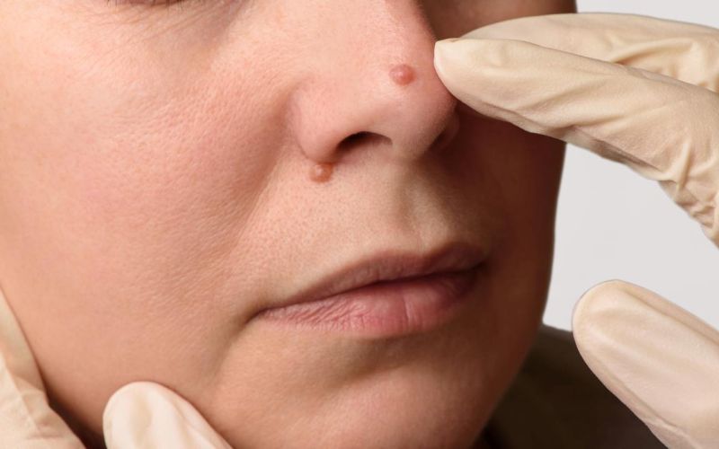
Moles are very common skin lesions. Most of them are small and do not pose an aesthetic problem for patients. Few people pay attention to them, and some even think they add charm. They are most often removed because of their excessive size and for health reasons - some moles may contain cancer cells and may even transform over time into melanoma, a malignant skin cancer. When should a pigmented mole catch our attention, and what does its removal involve? An increasingly popular, less invasive alternative to the scalpel is laser mole removal in Wrocław.
Table of contents
What are moles?
"Moles" is the common name for melanocytic naevi, which can occur all over the body - most often on the limbs, trunk and face. On average, each of us has about 20 such moles. Their appearance varies greatly in size, colour, shape and surface structure. Most moles are benign - they are small, brown, round and flat spots that do not attract particular attention. An aesthetic, and often also a health, problem arises with deviations from this norm, e.g. when a mole is large, asymmetrical and unevenly coloured.
How do moles form?
Moles form as a result of the proliferation (multiplication) of melanocytes, the cells responsible for producing melanin - the pigment that gives colour to the skin, eyes and hair. Depending on when they appear, pigmented lesions are divided into:
- congenital - present at birth, relatively rare (estimated to occur in 1% of newborns), ranging in diameter from a few millimetres to as much as a dozen or more centimetres (sometimes covering large areas of the body),
- acquired - they develop over time, mainly as a result of UV radiation. They can be further divided into benign (oval, small, brown spots) and atypical (asymmetrical, larger, with uneven colour - these are the ones that carry a risk of developing melanoma).
Which moles should you pay attention to?
The ABCDE method helps with self-assessment of worrying features of moles. It focuses on 5 main aspects; if you notice any of them, you should see a doctor. They are:
- A - asymmetry (in English: asymmetry) - deviations from an oval shape,
- B - border (in English: border) - irregular, ragged edges,
- C - colour (in English: color) - uneven colour, e.g. a distinct ring around the mole,
- D - diameter (in English: diameter) - size over 5 mm,
- E - evolution (in English: evolution) - progressive changes, e.g. enlargement of the mole.
This list does not cover the full spectrum of features that deviate from the norm and indicate disease. A medical consultation is also necessary if you feel pain, burning or itching at the site of the lesion, and if moles bleed or produce discharge.
Surgical mole removal - description of the procedure
Mole removal is a surgical procedure performed with a scalpel under local anaesthesia. In addition to the mole itself, the doctor also removes a small margin of unchanged tissue, which could potentially contain diseased cells; stitches and a dressing are then applied. As standard, the stitches are removed 7 days after the procedure, but this time may be longer depending on the type of mole removed and the course of healing.
This procedure must be preceded by a medical consultation, during which the doctor assesses the lesion and the patient's health and, if necessary, orders additional tests.
As with any procedure of this type, proper care of the operated area and following the doctor's recommendations are important. Above all, you should avoid baths and other activities that expose the wound to prolonged soaking. Exposing the operated area to the sun is also not advisable - it can lead to scarring and disrupt the healing process.
You can find more information about the procedure here: surgical mole removal - Wrocław.
Can moles be removed with a laser?
Some pigmented moles can be removed with a laser. However, this method is not used for diseased lesions. If, during the consultation, the doctor finds a risk of the mole becoming malignant, they will recommend a surgical procedure, which allows further histopathological examination.
—
Sources:
[1] Włodarkiewicz A., 2009, Znamiona melanocytowe wrodzone [Congenital melanocytic naevi], Przegląd Dermatologiczny 96
[2] Hałas P., 2017, Charakterystyka czerniaka oraz ocena poziomu wiedzy studentów na temat czerniaka złośliwego [Characteristics of melanoma and assessment of students' knowledge of malignant melanoma], Dermatologia
[3] Zaal L.H. et al., 2004, Classification of congenital melanocytic naevi and malignant transformation: a review of the literature
[4] Damsky W., Bosenber M., 2017, Melanocytic nevi and melanoma: Unraveling a complex relationship, Oncogene 36(42)