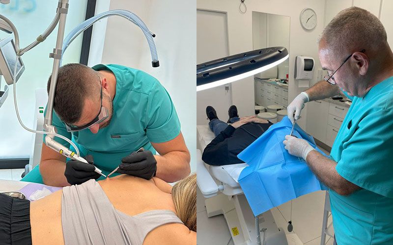
Skin lesions can develop as a result of various factors. Primary lesions, such as macules, papules, nodules, pustules or blisters, are usually the result of a developing disease. They are in turn followed by secondary lesions, e.g. scars, erosions and excoriations. After identifying the type and cause of the lesions, the doctor decides whether to remove them. There are many methods of performing such a procedure, and the most popular include laser and surgical removal. How do they differ?
Table of contents
When should skin lesions be removed?
Removing skin lesions is most often part of treating a disease or a preventive measure taken, for example, to prevent the development of cancer. However, even when a lesion poses no threat, a patient may decide to have it removed for aesthetic reasons, especially when it is in a visible place, e.g. on the face.
In practice, any worrying lesion should be checked by a doctor. What should particularly concern the patient? The ABCDE method developed by the American Cancer Society can help here. According to its principles, 5 features may indicate that a lesion is cancerous:
- A – asymmetry (one side differs from the other),
- B – borders (irregular shape),
- C – colour (uneven colouring),
- D – diameter (the lesion is larger than 5 mm),
- E – evolution (rapid change in colour, size, shape or thickness).
Differences between laser and surgical removal of skin lesions
Surgical removal of skin lesions involves cutting out the lesion with a scalpel. This method is used for large skin lesions (usually over 1 cm in diameter) and for precancerous or cancerous lesions. The excised skin tissue can undergo histopathological examination, which is used for further diagnosis.
Laser removal of skin lesions, on the other hand, involves removing the lesion with a laser beam whose light is absorbed by water, leading to vaporisation of the skin lesion. Contraindications to this procedure may include cancer, skin infection at the site of the lesion, taking anticoagulants, as well as pregnancy and breastfeeding.
The laser method works particularly well for benign lesions such as fibromas - see laser fibroma removal in Wrocław.
The table below shows the basic differences between surgical and laser removal of skin lesions.
Table 1. Comparison of surgical and laser removal of skin lesions
| Feature | Surgical procedure | Laser procedure |
| Most common types of lesions | pigmented moles, inflammatory nodules, cancerous and precancerous lesions | warts, fibromas, milia, xanthelasma |
| Duration of the procedure | approx. 15-30 minutes | approx. 5-30 minutes |
| Anaesthesia | local | local (an anaesthetic ointment is often sufficient) |
| After the procedure | After excising the lesion, the doctor places stitches, which are removed after approx. 7-14 days | After the laser procedure, a small scab forms at the site of the skin lesion |
| Recovery | 7-21 days | 7-10 days |
| Scar | size and visibility of the scar depend on the procedure; usually small and barely visible | no scar or a small scar (less visible than after surgical removal) |
PLEASE NOTE! The data in the table are approximate and may vary depending on each patient's individual case.
Laser or surgery? Choosing a method of removing skin lesions
It is not possible to state definitively which method of removing skin lesions is better. The doctor decides on the treatment method, taking into account the type and size of the lesion, as well as its location. Factors such as contraindications to the procedure, side effects and potential risks for the patient are also important.
One of the issues that interests patients most in connection with the removal of skin lesions is the visibility of the scar after the procedure. Some studies indicate that the choice of method is of little significance for the aesthetic results. One example is a comparison of the effectiveness of surgical, electrosurgical and laser treatment of benign moles carried out by researchers from India [1]. It turned out that in the long term (after 90 days), the differences in wound healing and scar appearance were not statistically significant for any of the three methods used. However, if the scar after the procedure is large and very visible, the patient may decide to have it removed with a laser at a later time.
—
Sources:
[1] Reddy Bandral M, Gir PJ, Japatti SR, Bhatsange AG, Siddegowda CY, Hammannavar R., A Comparative Evaluation of Surgical, Electrosurgery and Diode Laser in the Management of Maxillofacial Nevus, J Maxillofac Oral Surg. 2018